Record-Breaking Magnet Decodes Complex Molecules
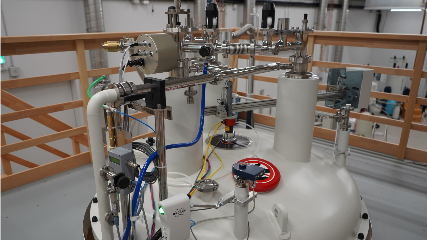
The strongest available permanent magnet is held in a container reminiscent of an oversized thermos flask. At just over 4 meters high, weighing more than 8 tonnes and resting on three stable supports, it is a solid symbol of hope for researchers, who use it to find out how highly complex biomolecules form and behave. On the inside, more than 1,800 liters of liquid nitrogen and helium keep the magnetic coils of superconducting wire at minus 271 degrees Celsius.
That is the only way to generate the enormous field strength of 25.8 teslas, and the stronger the magnetic field, the higher the resonance frequency at which the atomic nuclei of hydrogen, carbon and other elements respond when excited by radio waves (see box on nuclear magnetic resonance spectroscopy below). This extra power helps the Zurich experts out of a quandary: the larger and more complex the molecules, the more signals their protons deliver. With space on the spectra for these signals limited, things can get crowded. Sometimes the amplitudes overlap so much that it is hard to tell them apart, and thus to interpret them.
How an NMR spectrometer works
This type of spectrometer employs the principle of nuclear magnetic resonance. The instrument's main component is a very strong magnet. If you introduce a sample such as a liquid with molecules dissolved in it, into this magnetic field, the atomic nuclei align themselves with the field like small compass needles, because they have a spin like hydrogen or carbon nuclei.
To allow measurements to be taken, the instrument then targets the sample with radio waves, causing the atomic nuclei to change direction for a time. They subsequently return to their original state, releasing energy in the form of radio waves. The NMR spectrometer captures and measures these waves as signals from which a computer calculates an NMR spectrum. These spectra are often very complex if the molecules are large. From the sequence and amplitude of these numerous signals, researchers can identify the atoms’ chemical environment and the distance between them. This ultimately allows them to determine how a molecule is structured.
The new spectrometer has a distinct advantage over earlier models. Higher resonance frequencies mean clear separation between the countless signals that the instrument delivers from its readings. The spectrum becomes easier to read, not least because the greater magnetic field allows genuine signals to be distinguished better from the inevitable interference, or “noise”, which is one of the fundamental problems of this measuring technology.
Versatile tool for researchers
The new nuclear magnetic resonance spectrometer is, in essence, a high-tech magnifying glass for biomolecules in liquid samples. Its improved resolution could open up whole new perspectives on, say, fundamental biochemical research, the medical sciences, or pharmacology, as well as fields that we can barely imagine today.
The CHF 13.5 million needed to acquire the spectrometer was raised jointly by UZH, ETH Zurich and the University of Basel (which did much of the preparatory scientific work) under a license agreement withing the Swiss High-Field-NMR Facility. “I saw it as a really sensible way of financing such an expensive research instrument,” says chemist Oliver Zerbe, co-head of the Nuclear Magnetic Resonance (NMR) team, “and it ultimately means we will get the most out of it.”

The resolution delivered by this spectrometer is just much higher than previous instruments. It's like having a bigger chip in a camera.
Researchers are also conscious of economy in their day-to-day work. For example, they have significantly reduced the volumes of helium, a costly coolant, that they need to buy in by installing their own evaporated helium recovery system. Partners at the ETH then turn the helium back into a liquid and return it to the NMR facility to keep the spectrometer running less expensively over the long term.
Deployed in a wide range of fields
The Zurich researchers are optimistic that the 1.2-gigahertz spectrometer will deliver manifold new findings, Zerbe explains. “The resolution delivered by this spectrometer is just much higher than previous instruments. It's like having a bigger chip in a camera,” he says. “That makes it easier for us to see how substances bond to large, complicated molecules in bacterial cells.”
Antibiotic resistance is one example here. Zerbe's team is working on a natural substance called thanatin, derived from insects. It has active agent potential because it prevents bacteria from constructing their envelopes from two membranes. This bond is too unstable in human blood serum, however. By using NMR spectroscopy the researchers were able to pinpoint where and how thanatin blocks the formation of a molecular bridge between the membranes, thereby preventing bacteria from replenishing the nutrients they need.

You can think of intrinsically disordered proteins like spaghetti.
Under these conditions they were able to manipulate the molecule so that it remains stable in the blood, while producing less resistance and still maintaining its antibiotic effect. Zerbe continues: “In the past optimizations like these demanded attempt after attempt. We were basically flying blind.”
Decoding proteins with flexible structures
The new NMR technology allows processes to be planned more closely and executed more precisely. This is a huge advantage, for fundamental research, too. In the past, scientists mainly concentrated on complex molecules with fixed structures. This has broadened to include compounds that experts find fascinating because of their flexibility: intrinsically disordered proteins, or IDPs for short. These have no fixed structure and are largely made up of flexible chains instead.
“You can think of them like spaghetti,” says Ricarda Törner, assistant professor and NMR specialist at the Department of Chemistry. These compounds can adopt a whole variety of structures to fulfill different functions, whether to transmit signals or to regulate the processes involved in cell division. It is these vital processes that Törner is exploring with her team.
She explains: “Before a cell decides to divide, it needs external signals. These are answers to questions such as ‘are there enough nutrients?’ or ‘is our DNA actually intact?’” IDPs often take on this messaging function. Offering sharper resolution for molecules in liquid samples, the spectrometer is currently the best way of understanding these highly complex polymers and how they work. In her group, Törner is additionally working on new options for “labeling” these proteins chemically, in other words modifying them specifically to replicate states like that inside a cell in a test tube. For example, she might attach groups of phosphates at certain points and then use NMR spectra to examine what structural changes are triggered.

Törner offers the p53 molecule as a high-profile example of the importance of IDPs. Research groups around the world are working on this protein, which contains long, intrinsically disordered sections. In its normal state it prevents cells with faulty genetic information from dividing, and therefore provides a protective mechanism that can also extend to cancer. If p53 is damaged by mutations, tumors may result. Consequently, molecular researchers are looking for ways to manipulate it so that its function is restored.
Great hope for the future
The proteins that Törner is researching could also help us to better understand the process of cell division, which is an important element of tumor-related diseases. “In the near future we would like to study our target proteins in such depth that we properly understand their different states and can manipulate them very specifically,” she says. And in the long term? “We hope to understand the laws behind the processes and even to apply them universally one day.”
In the end, new findings like these could help to develop new therapies in the future, whether discovering alternative types of antibiotic, or identifying new treatment pathways for cancer.